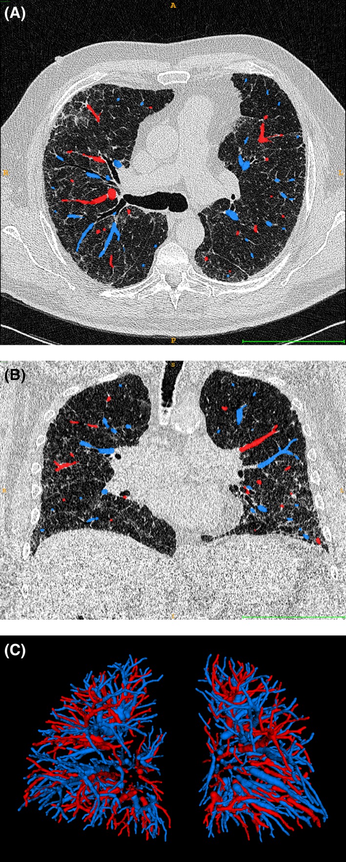
Figure 1.

Transversal (A) and coronal (B) thoracic computed tomography (CT) images of a representative patient with overlays of the arteries (blue) and veins (red) and 3D rendering of the arterial and venous vessel trees in this patient (C). The bar at the bottom of (A) and (B) is 10 cm wide.
