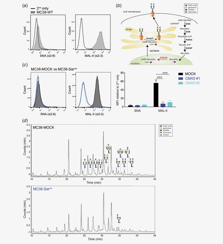
Figure 1.

Cell surface sialic acid expression is abrogated in the CMAS gene knockout. (a) The presence of α2‐6 and α2‐3 Sia in MC38‐WT cells was assessed by flow cytometry using the plant lectins SNA and MAL‐II, respectively. (b) Schematic representation of the sialylation pathway. (c) Two CRISPR/Cas9 guideRNAs targeting distinct regions in the CMAS gene both completely abrogate MC38 sialylation (MC38‐Sianull) as measured with plant lectins SNA and MAL‐II. MOCK‐transfected MC38 cells (MC38‐MOCK) were used as a negative control. Relative mean fluorescence intensities (MFI) were calculated relative to MFI of the 2nd antibody only. Data are representative of three independent experiments (a and c). Mean ± SD; ***, p < 0.001. (d) Liquid Chromatography‐Mass spectrometry (LC–MS) analysis of MC38‐MOCK (upper) and MC38‐Sianull (bottom) sialylation. Only representative examples of the identified sialylated glycans structures are depicted. [Color figure can be viewed at wileyonlinelibrary.com]
