Figure 1.
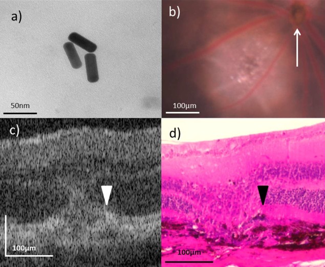
GNR functionalization and imaging. (a) Transmission electron micrographs of GNR demonstrating unaltered morphology following surface functionalization. (b) In vivo fundoscopy showing a day 5 LCNV lesion adjacent to the optic nerve head (white arrow). (c, d) Representative OCT in vivo and hematoxylin and eosin stained ex vivo images of day 5 LCNV lesions, with disrupted RPE (white and black arrowheads, respectively) present.

