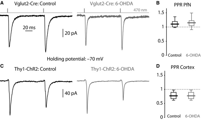
Figure 2.

Optogenetic PPRs in ChIs from control and dopamine‐depleted striata. (A) Averaged EPSCs evoked in ChIs by a pair of 470‐nm light pulses (100‐ms inter‐pulse interval) that activate ChR2‐laden PfN fibers from a control (left) and 6‐OHDA‐lesioned (right) Vglut2‐Cre mouse. (B) Boxplot of PPRs at PfN synapses onto ChIs in control and 6‐OHDA‐lesioned mice. (C) Averaged EPSCs evoked in ChIs by a pair of 470‐nm light pulses that activate ChR2‐laden fibers from a control (left) and 6‐OHDA‐lesioned (right) Thy1‐ChR2 mouse. (D) Boxplot of PPRs at nominally cortical synapses onto ChIs in control and 6‐OHDA‐lesioned mice.
