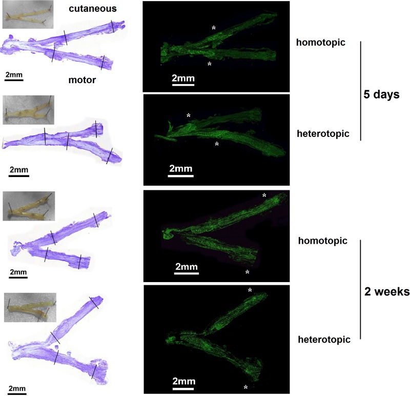
FIGURE 4.
Representative examples of histological evaluation after homotopic/heterotopic nerve grafting. The proximal and distal coaptation sites in the motor as well as the cutaneous nerve can be seen in the macroscopic picture taken directly after excision. Suture sites are indicated in the cresyl violet overview (left panel). Axonal regeneration was visualized by Neurofilament 200 staining (right panel). Axonal regeneration front is staggered at the proximal coaptation site at 5 days (highlighted by asterisks). Two weeks after repair, axonal regeneration has surpassed the distal coaptation site in both grafting modalities.

