
Figure 5.
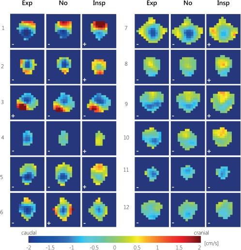
CSF net flow for all aqueduct voxels during expiration gating (Exp), inspiration gating (Insp), and without respiratory gating (No) for all 12 subjects, for the first measurement. For most subjects, during expiration gating most voxels show caudal net CSF flow, and during inspiration gating more voxels show (larger) cranial net CSF flow. The net CSF flow directions are indicated with ± symbols for cranial/caudal net CSF flow.