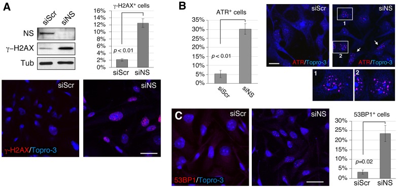
Fig. 1.
Loss of nucleostemin increases the number of DNA damage foci as visualized by staining for multiple DNA-damage-response proteins. MDA-MB-231 cells were treated with scrambled (siScr) or nucleostemin-specific (siNS) siRNA duplexes for 48 hours at 100 nM. (A) Upper left panel, western blots of nucleostemin and γ-H2AX. Protein loading was controlled by the amount of α-tubulin (Tub). NS, nucleostemin. Lower panels, immunofluorescence with an anti-γ-H2AX antibody. Upper-right panel, quantitative measurement of the percentage of γ-H2AX+ cells in siScr- and siNS-treated samples. (B) Percentage of ATR+ cells (left) as determined by using immunofluorescence (right). Panels 1 and 2 show magnified views of the regions outlined in white. White arrows, examples of ATR+ cells. (C) The percentage of 53BP1+ cells (right) as determined by using immunofluorescence (left). Scale bars: 50 µm (A and C), 20 µm (B). Bar graphs represent mean±s.e.m.

