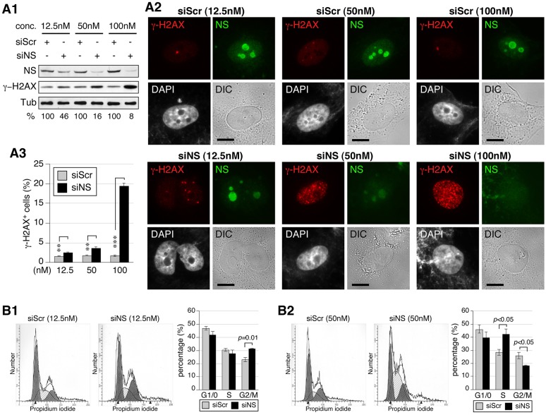
Fig. 4.
The knockdown efficiency dictates the nature of the cell-cycle arrest of nucleostemin-depleted cells. (A1) Western blots to show the knockdown efficiency in MDA-MB-231 cells treated with different concentrations (conc.) of siScr and siNS. Samples were collected at the 48-hour time-point. Relative nucleostemin intensities (%) between paired siScr and siNS samples are shown below the blots. α-tubulin (Tub) is shown as a loading control. NS, nucleostemin. (A2) Immunofluorescence performed using anti-nucleostemin and γ-H2AX antibodies in siScr- and siNS-treated MDA-MB-231 cells. Nuclear structure and cell morphology are shown by DAPI staining and differential interference contrast (DIC) imaging, respectively. Scale bars: 10 µm. (A3) FACS-based quantification of the percentage of γ-H2AX+ cells in siScr- and siNS-treated samples. **P<0.001, ***P<0.0001. (B) Cell-cycle profiles and quantitative analyses (n = 4) of MDA-MB-231 cells treated with 12.5 nM (B1) or 50 nM (B2) siRNAs for 48 hours. Data show the mean±s.e.m. of three independent experiments.

