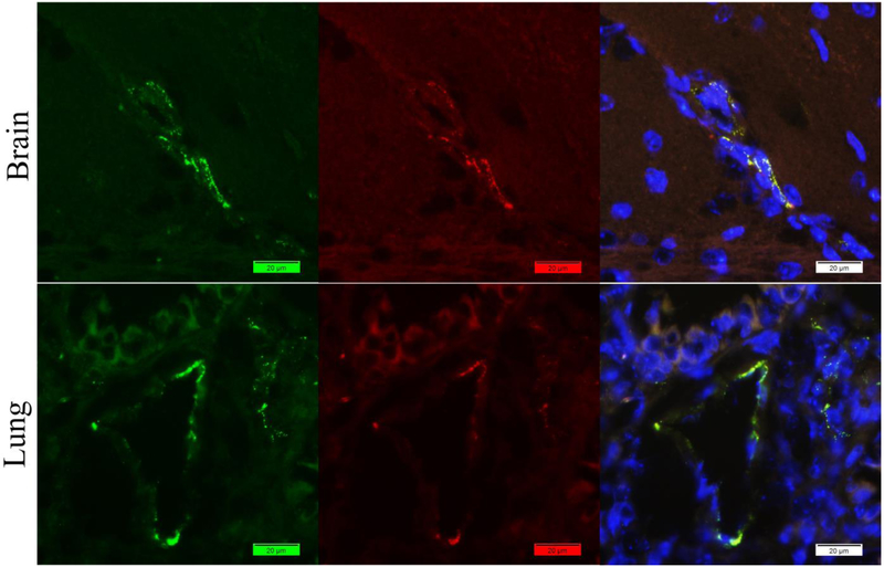
Figure 4.
Staining of mouse brain and lung tissues with rabbit anti-vWF antibody (Green) and rabbit anti-fibrin(ogen) antibody (Red) by our new method but without normal rabbit serum. Promising vWf signals in brain and lung. However, red color shows similar distribution with the green ones which indicated the labeled rabbit anti-fibrin(gen) antibody have bounded with the free binding sits of previous antibodies.

