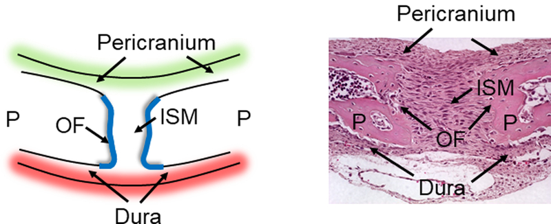
Figure 2.

Schematic and histological presentation of the sagittal suture. (A) A schematic and (B) histological appearance of the sagittal suture showing the paired parietal bones (P) and the relative positions of the osteogenic fronts (OF), intrasutural mesenchyme (ISM), the pericranium and the dura mater. The leading edges of these osteogenic fronts contain proliferative osteoprogenitor cells and the sagittal suture is a composite structure that consists of the osteogenic fronts and the intrasutural mesenchyme.
