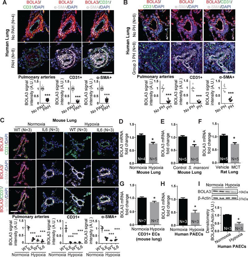
Figure 1: Down-regulation of BOLA3 across multiple in vitro and in vivo models of PH.
(A-C) Fluorescence microscopy of human lung from Group 1 PAH (A) and Group 3 PH (B) vs. non-PH patients; interleukin-6 (IL-6) transgenic mice (without or without hypoxia); and hypoxic wildtype (WT) mice (C) revealed reduced BOLA3 expression in endothelium (CD31 label) and smooth muscle (α-SMA label). Scale bar: 50μm. (D-G) By RT-qPCR, BOLA3 transcript expression was decreased in lung from hypoxic PH mice (10% O2) (D), PH mice infected with S. mansoni (E), and monocrotaline (MCT)-exposed PH rats (F). (G) Similarly, BOLA3 transcript expression was decreased in CD31-positive pulmonary endothelial cells (ECs) isolated from chronically hypoxic PH mice. (H-I) By RT-qPCR and immunoblotting respectively, BOLA3 was decreased at the transcript (H) and protein (I) levels in cultured hypoxic human pulmonary arterial endothelial cells (PAECs). In (I), representative immunoblot is shown with densitometry calculated across 3 separate replicates. In all panels, mean expression in controls (no PAH, no PH, WT, vehicle, control, normoxia) was normalized to a fold change of 1, to which relevant samples were compared (N = 3–8/group). Data represent the mean ± SEM (*P < 0.05, ***P < 0.001).

