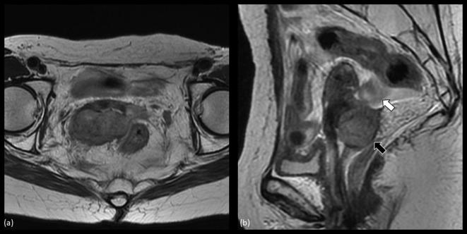
Figure 1.
MRI of recurrent cervical cancer before salvage brachytherapy. (a) shows an axial image of the tumor. The tumor extended beyond the right-sided parametrium to the pelvic wall. (b) depicts a sagittal image of the tumor (black arrow) visualizing that the sigmoid colon (white arrow) is located just next to the recurrent lesion.

