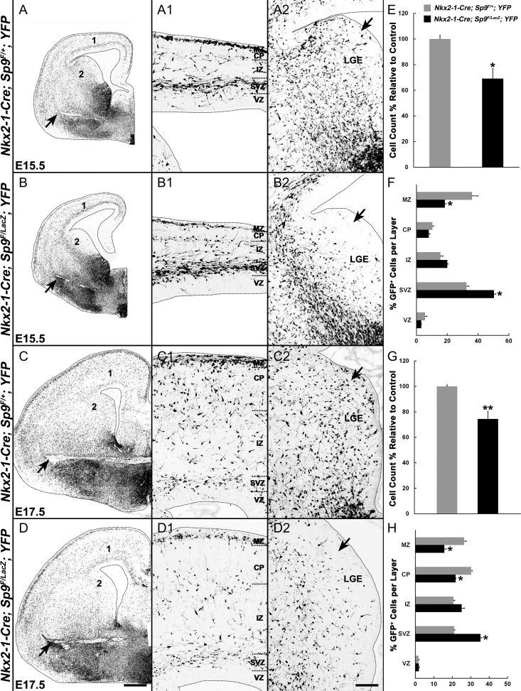
Figure 3.
MGE-derived interneurons are reduced and distributed abnormally in the cortex of Sp9-CKOs. (A–D2) GFP immunostaining on coronal hemisections showing the distribution of MGE-derived cells (GFP+) in telencephalic hemispheres at E15.5 and E17.5. Note that GFP+ cells in the LGE VZ/SVZ of mutants were absent or greatly reduced at E15.5 and at E17.5 (arrows in A2–D2). More GFP+ cells were observed in the ventral telencephalon of Sp9-CKOs (arrows in A–D). (E–H) Quantification showing that Sp9-CKO mice had reduced numbers of GFP+ cells in the cortex at E15.5 (E) and E17.5 (G). The percentage of GFP+ cells was reduced in the marginal zone (MZ) and cortical plate (CP) (F, H), and increased in the SVZ (F, H). IZ, intermediate zone. Student’s t-test (E, G) and χ2 test (F, H), *P < 0.05; **P < 0.01, n = 3. Scale bars: 200 μm in D for A–D; 100 μm D2 for A1–D2.

