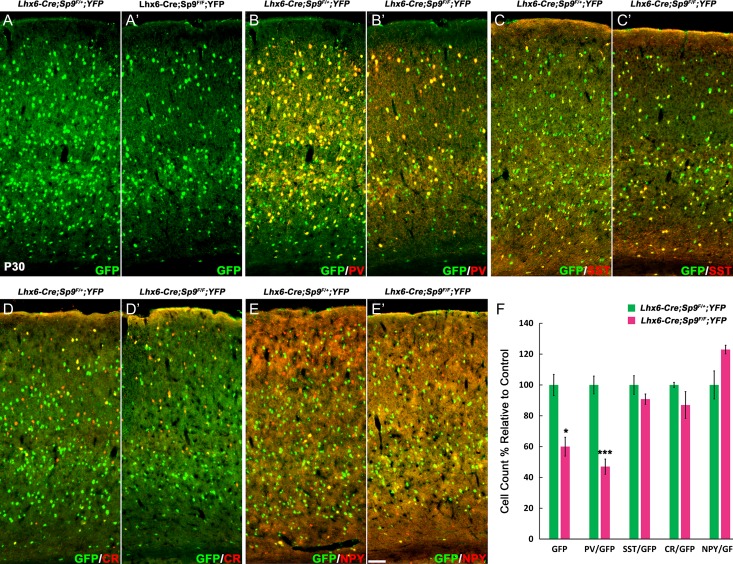
Figure 5.
Cortical interneurons are reduced in Lhx6-Cre; Sp9F/F; Rosa-YFP mice. (A–E′) GFP and interneuron markers double-immunostaining on P30 cortex coronal sections. (F) Quantification of above experiments. GFP+ and GFP+/PV+ cells were significantly reduced. Student’s t-test, *P < 0.05; **P < 0.01, n = 3. Scale bar: 100 μm.

