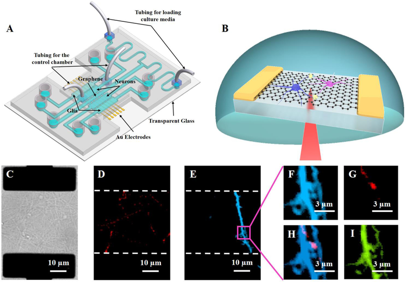
Fig. 1.
(A) Schematic of a four-chamber neuron-glia co-culture microfluidic device with integrated graphene transistors. (B) Schematic of scanning photocurrent measurements. A diffraction-limited laser spot passes through a transparent coverslip to scan over the graphene underneath neurons. (C) Differential interference contrast (DIC) of a graphene transistor underneath neural networks. The two black rectangles are opaque Au electrodes that are underneath the graphene membrane. Neurons, at day 5 in culture, were differentially transfected with (D) mCherry-synaptophysin (red) and (E) mCerulean (blue), maintained in co-culture with glia. Zoom-in fluorescence images of a magenta square region in Fig. 1E: (F) mCerulean (blue); (G) mCherry-synaptophysin (red); (H) overlay of mCerulean and mCherry-synaptophysin; and (I) GFP-GCaMP6s (green).

