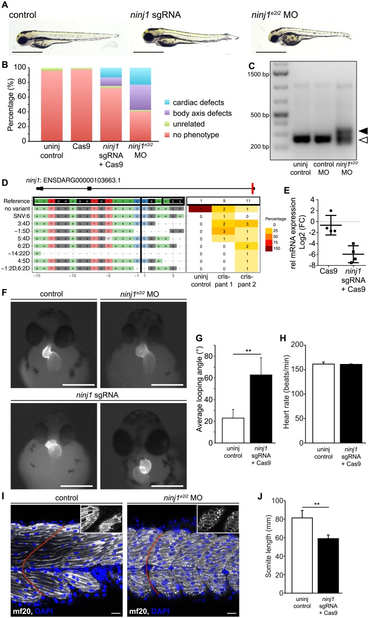
Fig 7. Ninjurin1 deficiency impairs zebrafish heart and skeletal muscle development.
Zebrafish (wild type or transgenic Tg(myl7:EGFP)twu34) embryos were injected with ribonucleoprotein complex of Cas9 and sgRNA targeting exon 1 of ninjurin1 (ninj1 sgRNA) or morpholino targeting exon2/intron2 junction of ninjurin1 (ninj1e2i2 MO) at one cell stage and compared to uninjected control. In all panels, embryos were analysed at 48–50 hours post fertilization (hpf). (A) Representative images of uninjected controls and ninjurin1-deficient embryos showing cardiac and body axis defects. Scale bar, 1 mm. (B) The relative occurrence of the cardiac phenotypes is depicted. (C) qRT-PCR of ninj1 cDNA from uninjected control, control morpholino injected (control MO), and ninje2i2 MO injected embryos at 50 hpf. In control MO injected embryos, the size of the ninj1 amplified fragment is 254 bp (empty arrowhead), whereas it is approximately 350 bp in ninj1 morphants, due to defective splicing of intron 2 (full arrowhead). (D) Panel plot showing allele variations in ninj1 sgRNA-Cas9-injected embryos compared to the WT ninj1 allele (no variant) (ENSDARG000001036663.1). (E) Relative mRNA expression of the ninj1 gene in uninjected, Cas9, and ninj1 sgRNA-Cas9-injected embryos by qRT-PCR and normalized to eef1α1l mRNA expression. Data represent the log2 fold change of ninj1. Cas9 to uninjected control, ns, P = 0.0686, ninj1 sgRNA-Cas9-injected embryos to uninjected controls, **, P = 0.0025. (F) Representative images of Tg(myl7:EGFP)twu34 showing defects in cardiac looping and dextrocardia in ninj1-deficient embryos. Scale bar, 1 mm. (G) Quantification of the looping angle, ninj1 sgRNA-Cas9-injected embryos to uninjected control, **, P = 0.0012. (H) Heart rate remained unchanged in ninj1-deficient embryos; ninj1 sgRNA-Cas9-injected embryos to uninjected control, ns, P = 0.8406. (I) Single confocal plane of whole-mount embryos showing skeletal muscle cells and their nuclei stained with anti-myosin heavy chain antibody (mf20) and DAPI, respectively. Myosin assembles within the sarcomeres in controls, while it forms aggregates in ninj1-deficient embryos (in insets, apical section). Somites do not display the typical chevron shape, red line. Scale bar, 20 μm. (J) Somite length was significantly shorter in ninj1-deficient embryos compared to controls; **, P = 0.0025.

