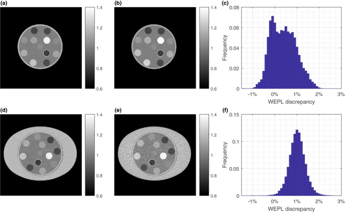
Figure 5.

(a) and (b) show the SPR images of the head phantom reconstructed by the JSIR‐BVM method and the image‐based HS methods, respectively. (c) shows the distribution of the differences between WEPL computed from the two SPR images of the head phantom. (d) and (e) show the SPR images of the body phantom reconstructed by the JSIR‐BVM method and the image‐based HS methods, respectively. (f) shows the corresponding WEPL difference for the body phantom. The WEPL is computed for 150 parallel beams with width of 1 mm for every degree. [Color figure can be viewed at wileyonlinelibrary.com
