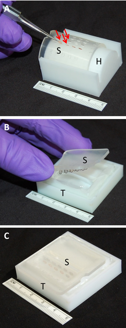
Figure 2.

A) Leica EM AC20 grid support plate (S) stretched out to open slits and clipped into modified Hiraoka plate holder (H) with TEM grids (arrows) loaded using fine tipped forceps. B) Grid support plate (S) containing TEM grids placed into staining solution within tray (T) at an angle to avoid bubble formation. C) TEM grids in support plate (S) submerged in stain filled in tray (T.)
