
Figure 3.
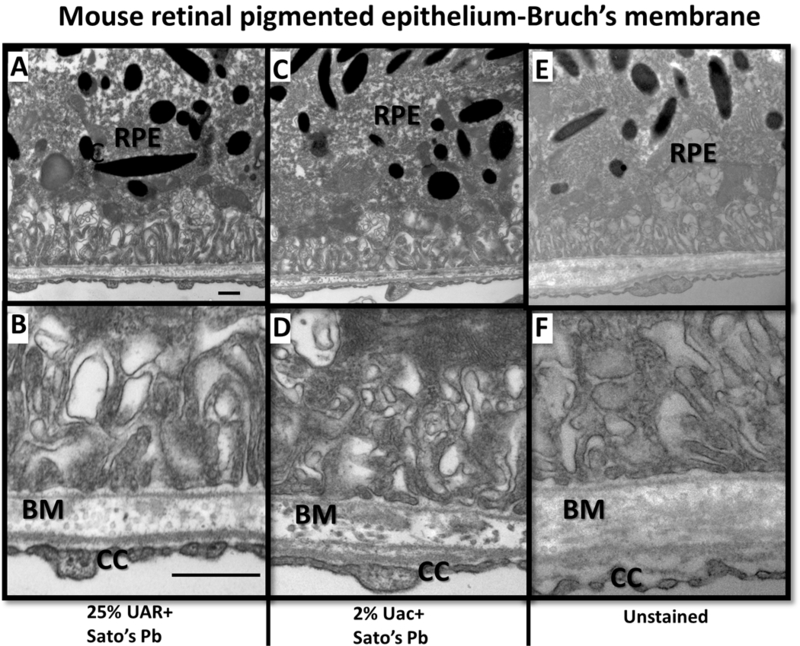
TEM of individual mouse eye sample from set A (without Uac en bloc staining). Retinal pigmented epithelium (RPE) cells and Bruch’s membrane (BM) stained with 25% UAR+Sato’s Pb stain (A & B), 2% Uac+Sato’s Pb stain (C&D) compared to unstained thin section (E&F.) Higher magnification images (B, D, E) of basal RPE and BM interface with surrounding fenestrations of choriocapillary (CC). BM matrix is electron dense in the dual stained 25% UAR+Sato’s Pb stain (A & B), 2%Uac+Sato’s Pb sections. Images A, C, E acquired at 18,500x magnification, scale bar =500 nm. Images B,D,F acquired at 30,000x magnification, scale bar = 500nm.