Figure 2.

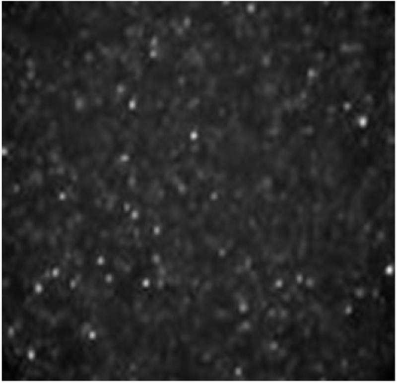
Fluorescent image of liposomes in alginate. The image is a representation of a z-section. As can be seen, a relatively homogenous distribution of liposomes is contained within each microbead. Microbeads were 200 μm +/− 5% in diameter.

Fluorescent image of liposomes in alginate. The image is a representation of a z-section. As can be seen, a relatively homogenous distribution of liposomes is contained within each microbead. Microbeads were 200 μm +/− 5% in diameter.