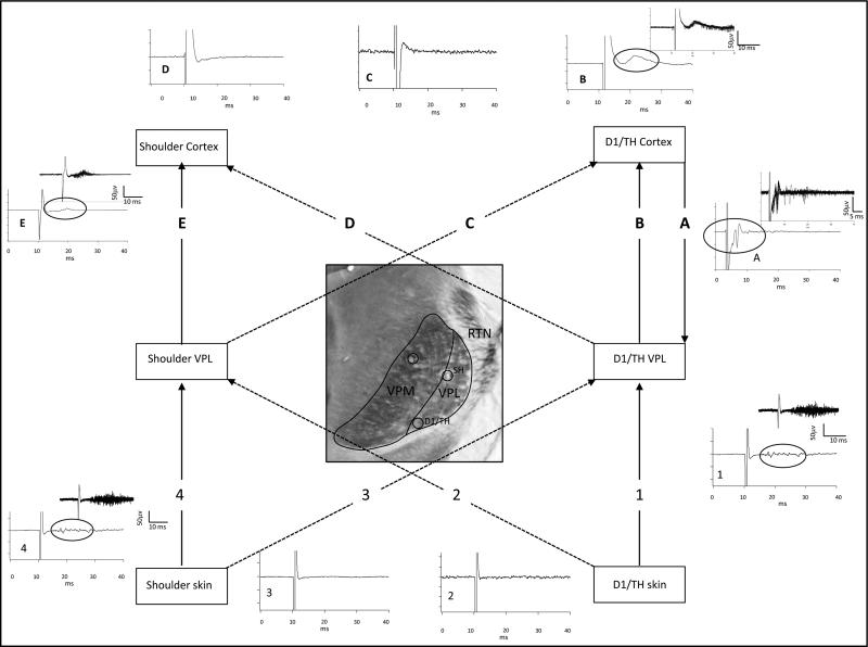
Fig. 3.
Pattern of projection from digit and shoulder periphery to VPL (nos. 1–4) and from VPL to SI cortex (letters “A”–“E”). Locations of D1/TH and shoulder representations in VPL are shown in the photomicrograph of CO-stained VPL that were identified by stimulating the digit and shoulder skin surface as described in Fig. 2. Stimulation of D1/TH evoked a response in D1/TH region in VPL (solid line labeled “1”) but did not evoke a response in shoulder sites (dashed line labeled “2”) in VPL. Similarly, stimulation of the shoulder skin evoked a response in a homotopic shoulder region in VPL (solid line labeled 4) but did not evoke a response in the digit region in VPL (dashed line labeled “3”). Averaged traces are shown for each of the stimulating and recording sites. Traces 1 and 4 show averaged evoked responses for 10 consecutive stimulations; circles in the trace indicate the evoked response part of the record. Insets show a response buffer for these traces. Locations of the D1/TH and shoulder representations were also identified in SI. The recording electrodes in VPL were then used to deliver single-pulse stimulation and responses were examined at digit and shoulder locations in SI (letters “A”–“E”). Stimulation at the digit location in VPL evoked a response in the homotopic digit location in SI (solid line labeled “B”) but not in shoulder location in SI (dashed line labeled “D”). Stimulation at a shoulder responsive site in VPL evoked a response in the homotopic shoulder location in SI (solid line labeled “E”) but not in the D1/TH representation in SI (dashed line labeled “C”). Averaged traces are shown for each of these stimulation sites and buffers are displayed for those sites where evoked responses were recorded. The electrode in SI was then used for stimulation and the electrode in VPL was used for recording. An example of an antidromically-activated response is shown in trace “A”, but similar stimulation in shoulder SI failed to activate this digit site in VPL (data not shown).

