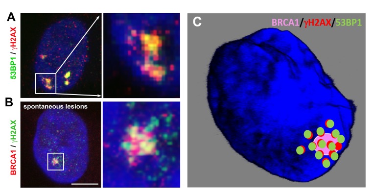
Figure 4.
(A) Nuclear arrangement of the 53BP1 (green) protein and γH2AX (red) in spontaneously occurring DNA lesions. (B) The nuclear distribution pattern of the BRCA1 protein (red) and γH2AX (green) in spontaneous DNA lesions. HeLa cells were used for these illustrations, which depict the primary results published by [67] and [20]. Scale bars, 10 µm. (C) A pictorial illustration of BRCA1/γH2AX/53BP1 compartmentalization at a spontaneous DSB site: Chapman et al. [67] showed a spatial link between 53BP1- and BRCA1-positive foci or 53BP1- and γH2AX-positive repair foci. Suchánková et al. [20] described the methodology of immunostaining and showed a degree of colocalization between 53BP1-γH2AX, 53BP1-MDC1, and MDC1-γH2AX.

