Figure 6.

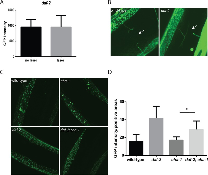
daf-2 mutations can protect C. elegans muscle from the effects of denervation or reduced cholinergic signaling. (A) In contrast to experiments performed in wild-type animals, the severing of motor neuron commissures via laser axotomy in daf-2 mutant animals did not reduce muscle mitochondrial mass in the denervated muscles as measured by fluorescence from a mitochondrial localized GFP. N >12 for both genotypes and treatments. (B) The differing effects were not simply due to the regrowth of the axons in the daf-2 mutants as both the wild-type (left image) and daf-2 mutants (right image) showed the presence of severed axons as indicated by the arrows. Both images were taken with confocal microscopy using a 100X oil-immersion objective. (C) The daf-2 mutation is able to rescue the effects of the cha-1 mutation on muscle mitochondrial structure. Shown are confocal images from adult day 15 wild-type, cha-1, daf-2, and daf-2 cha-1 mutants, expressing a mitochondrial-localized GFP to label the muscle mitochondria, grown at the permissive temperature 16ºC. These images show the age-related disruption of the filamentous mitochondrial structure in the wild-type animals which is reduced in the daf-2 mutants. Additionally, the chronic low-level disruption of cholinergic signaling in the cha-1 mutant even at permissive temperature exacerbates the disruption of muscle mitochondrial structure. However, the daf-2 mutation is also able to attenuate this decline in mitochondrial structure. (D) These declines can also be visualized by measuring the GFP+ area in the myocyte containing the mitochondria relative to the total muscle area in the confocal images. N >5 for all genotypes. * represents p < 0.05 by t‐test.
