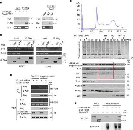
Fig. 2. PES1 forms an active complex with telomerase.

(A) MCF7 cells transfected with the indicated plasmids were immunoprecipitated with anti-Flag. The immune complexes were eluted with Flag peptide and re-IP using anti-Myc or normal IgG. The resulting precipitates were used for detection of Flag-hTERT and Myc-PES1 expression by immunoblot. hTR levels were determined by RT-PCR. (B) MCF7 cell extracts were fractionated on Superose 6 size exclusion columns. Fast protein liquid chromatography (FPLC) chromatographic elution profiles are indicated. The elution positions of calibration proteins with known molecular masses (kDa) are shown by arrows, and an equal volume from each chromatographic fraction was used for determination of protein expression by immunoblot with the indicated antibodies. To increase the specificity of hTERT detection, cells were immunoprecipitated with anti-hTERT from Abbexa, followed by immunoblot with anti-hTERT from Abcam (hTERT after pre-purification). hTR levels were determined by reverse transcription polymerase chain reaction (RT-PCR) and telomerase activity by telomerase repeat amplification protocol (TRAP). CHAPS buffer was used as a negative control for TRAP assay. IC, internal control; mAu, milli-Absorbance unit. Red frame indicates a potential complex with hTERT/hTR/DKC1/PES1. (C) MCF7 and U2OS cells were transfected with Flag-tagged PES1 or hTERT or empty vector, and immunoprecipitated with anti-Flag agarose. The resulting precipitates were used for assessment of Flag-PES1 or Flag-hTERT expression by immunoblot. hTR levels were determined by RT-PCR. β-Actin was used as a negative control for hTR determination. (D) MCF7-Flag-PES1+shPES1 cells or control (MCF7-Flag+shCon) cells were transfected with control small interfering RNA (siRNA) or hTERT siRNA as indicated and were analyzed as in (C). (E) Biotinylated hTR was incubated with purified GST-hTERT, GST-PES1, or GST-hTERT plus GST-PES1. The resulting complexes were subject to RNA pull-down assay. The amount of biotin-hTR was detected by the Chemiluminescent Nucleic Acid Detection Module Kit (Thermo Fisher Scientific).
