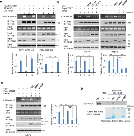
Fig. 3. PES1 enhances assembly of TERT and TR.

(A) PES1 WT or KO MCF7 cells (left) and MEFs (right) were transiently transfected with the indicated plasmids and immunoprecipitated with anti-Flag. The precipitates were used for assessment of Flag-h/mTERT, PES1, and β-actin expression by immunoblot. TR levels were determined by RT-PCR. Flag-h/mTERT, Flag-tagged human or mouse TERT. Flag-hTERT was used for MCF7 cells, and Flag-mTERT was used for MEFs. (B) siRNA-mediated PES1 KD MCF7 or HepG2 cells were transfected with Flag-hTERT and siRNA-resistant Myc-tagged PES1, PES1 (ΔBRCT), or PES1 (W397R) as indicated and were analyzed as in (A). (C) siRNA-mediated PES1 KD MEFs were transfected with Flag-mTERT and siRNA-resistant Myc-tagged PES1, PES1 (ΔBRCT), or PES1 (W397R) as indicated and were analyzed as in (A). Data shown are mean ± SD of three independent experiments (A to C). **P < 0.01. (D) Biotinylated hTR was incubated with purified GST-hTERT and WT or mutant His-PES1. The resulting complexes were subject to RNA pull-down assay. Unlabeled hTR was used as a negative control. Asterisks indicate the positions of the expected full-length fusion proteins.
