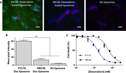
Fig. 5. Characterization of P1C10-targeted doxorubicin-loaded liposomes.

(A) VLR-targeted doxorubicin-loaded liposomes were used in a binding assay with murine brain tissue sections and imaged for doxorubicin fluorescence (green). GS-IB4 lectin labels microvessels (magenta), and cell nuclei are stained with Hoechst 33342 (blue). (B) Doxorubicin fluorescent signal, from groups presented in (A), is quantified for each group (*P < 0.01, ANOVA). (C) U87 cell viability as a function of doxorubicin concentration provided by VLR-conjugated doxorubicin-loaded liposomes bound to bEnd.3 ECM. The EC50 was determined by seeding U87 cells onto bEnd.3-derived brain ECM, incubating with VLR-conjugated doxorubicin labeled liposomes, washing unbound liposomes, and then culturing for 72 hours at which point the cell viability was measured. All dilutions were performed in triplicate. EC50 for P1C10 = 199.0 ± 1.7 nM and RBC36 = 3312.0 ± 2.6 nM (P < 0.05, Student’s t test).
