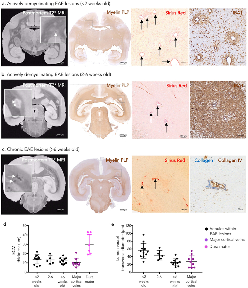
Figure 2.
MRI-pathology of venular remodeling in marmoset EAE lesion development Venular remodeling is seen early in marmoset EAE lesion development on both postmortem T2* MRI (white arrows) and histopathology. Magnified views of lesions with remodeled blood vessels (black arrows) are shown in the boxes.
(a) In lesions younger than 2 weeks, demyelination (myelin PLP) and inflammatory infiltrate (IBA1) are limited to the perivenular parenchyma.
(b) In lesions between 2 and 6 weeks old, both demyelination and inflammatory infiltrate are more extensive.
(c) Small demyelinated lesions older than 6 weeks (chronic) show prominent perivenular extracellular matrix deposition. Double staining of a representative venule for collagen IV (basement membrane, brown) and fibrillary collagen I (blue) from the same animal (non-contiguous paraffin section).
(d-e) Quantification (mean and standard deviation) of perivascular wall-thickness and lumen vessel transversal maximum diameter of venules within EAE lesions and major cortical veins showing ECM deposition. Remodeled venules within marmoset EAE lesions resemble major cortical veins in terms of size and presence of collagen perivascular deposition. See the text for statistical analysis.
Abbreviations: EAE: experimental autoimmune encephalomyelitis; myelin PLP: myelin proteolipid protein; ECM: extracellular matrix.

