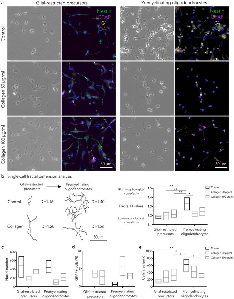
Figure 3.
- Glial-restricted precursors: in comparison to control, collagen I induced apoptosis (fragmented or pyknotic nuclei), bizarre cellular morphology (enlarged soma, loss of bipolar morphology, thick processes) and increased number of GFAP+/nestin- cells (b,c,d,e).
- Premyelinating oligodendrocytes: in comparison to control, collagen I induced lower cell counts (c), reduced ramification of cellular processes (b) and increased number of GFAP+/O4- cells (d).
(b) Single-cell fractal dimension (D) analysis. Representative cells at different differentiation stages, selected for the fractal dimension analysis, are shown with corresponding D values. D values quantify the degree of cellular morphological complexity (low in bipolar cells, high in ramified cells).
See the text for statistical analysis; Tukey’s post-hoc comparison *p<0.05; **p<0.01

