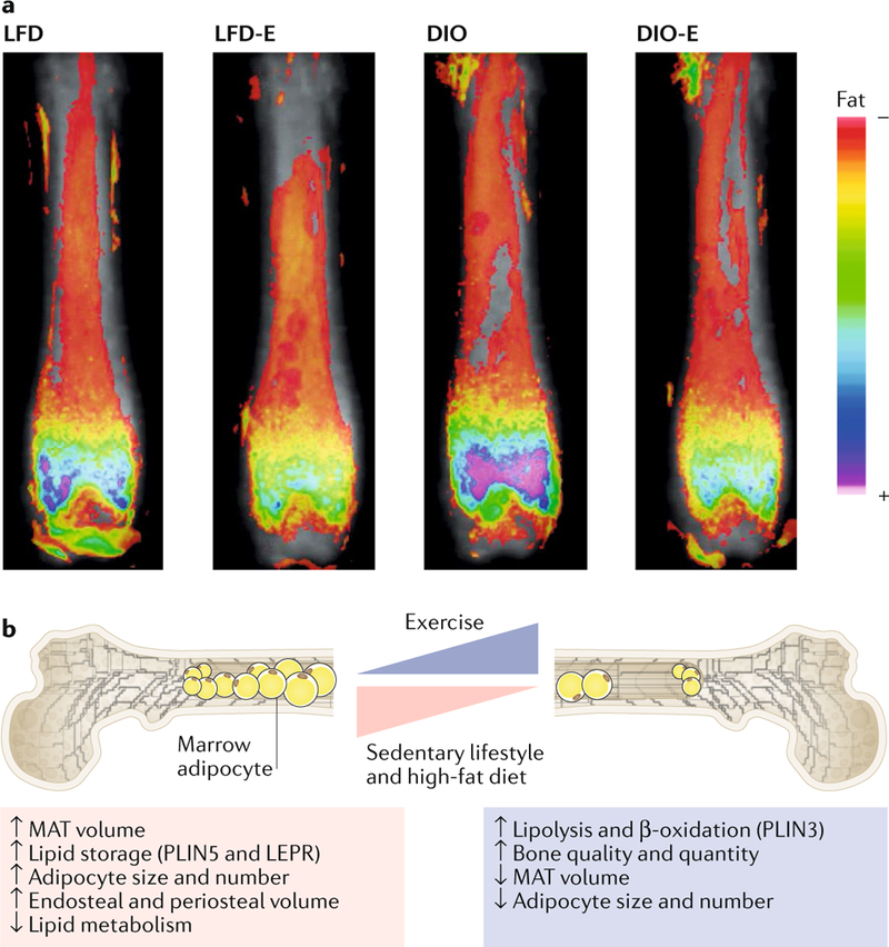
Fig. 2 |. Exercise suppresses expansion of marrow adipocytes and strengthens bone in obese mice.

a | Obese (diet-induced obesity (DIO)) and lean (low-fat diet (LFD)) mice were allocated to running exercise (DIO-E and LFD-E, respectively) or sedentary groups for 6 weeks (n = 6 per group). The images are a visualization of femoral marrow adipose tissue (MAT) in mice measured by MRI with advanced image analysis. Each image represents six images superimposed on each other. The heat map demonstrates the relative lipid quantity. b | Schematic representation of marrow adipocytes in the setting of obesity with or without exercise. DIO increases adipocyte size and number and expression of the lipid droplet marker PLIN5, resulting in expansion of cortical endosteal and periosteal bone surfaces. By contrast, exercise increases bone quantity and quality relying on β-oxidation of lipids in the marrow, as supported by a reduced number of adipocytes in the marrow and their cross-sectional area and increased expression of oxidation and lipolysis markers (for example, PLIN3). Part a reproduced with permission from REF.187,Wiley-VCH.
