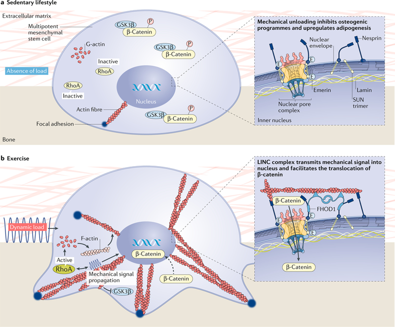
Fig. 3 |. Mechanotransductive responses of mesenchymal stem cells to dynamic mechanical stimuli are achieved through the internal stiffening of the cell via cytoplasmic-bound actin proteins.

a | The absence of mechanical forces prevents the polymerization of actin fibres, preventing the dephosphorylation of β-catenin, which remains bound to GSK3β. As such, β-catenin does not translocate to the nucleus, resulting in the expression of PPARγ-driven adipogenic pathways. b | By contrast, mechanical stimuli recruit actin fibres to the interface of the cell membrane and the substrate surface. These focal adhesions become stronger and denser in response to dynamic mechanical stimuli, permitting the movement of β-catenin into the nucleus and an ensuing osteogenic response. FHOD1, FH1/FH2 domain-containing protein 1; LINC, linker of nucleoskeleton and cytoskeleton.
