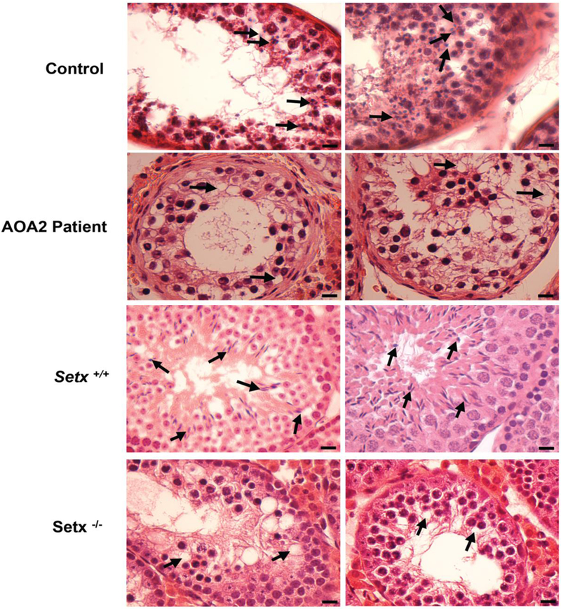
Figure 1. Spermatogenesis is disrupted in an AOA2 patient as shown by the absence of mature germ cells in seminiferous tubules.

Hematoxylin and eosin (H&E)-stained sections of testis from A. control and AOA2 patient sample, and B. adult Setx+/+ and Setx−/− mice. Black arrows indicate mature germ cells in the control and Setx+/+ while vacuolated seminiferous tubules in which both spermatozoa and mature spermatids are absent are observed in both AOA2 patient and Setx−/− seminiferous tubules. Scale bar, 20 μm.
