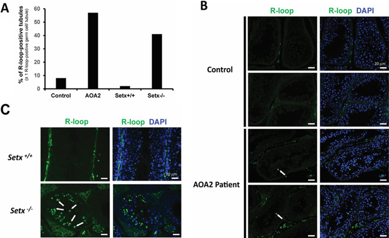
Figure 4. Detection of R-loops (DNA:RNA hybrid structures) in AOA2, control, Setx+/+ and Setx−/− testis.

A. Percentage of R-loop-positive tubules in Control, AOA2, Setx+/+ and Setx−/− samples. Tubules containing ≥ 1 R-loop-positive germ cell were scored as R-loop-positive tubule. B. Example of R-loop staining in control and AOA2 patient testis biopsy sections. C. R-loop staining in Setx+/+ and Setx−/− mouse testis seminiferous tubules. R-loops were detected in germ cells as shown by the white arrow. While many R-loop-positive germs cells were detected per tubule in Setx−/− mice as shown in (C), only one to two R-loop-positive germ cells were detected per tubule in the AOA2 patient sample (B).
