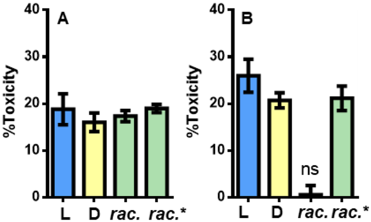
Figure 2.
Cytotoxicity of PSMα3 (5 μM total peptide) against HEK293FT cells as measured by an ATP-based luminescent viability assay. Rac. indicates preincubated in racemic form; rac* indicates L- and D- PSMα3 pre-incubated separately and mixed when added to cells. A: Peptides pre-incubated at 25 μM for 24 h before addition to cells. B: Peptides pre-incubated at 800 μM for 24 h before addition to cells. 0% and 100% toxicity represent averaged luminescence values of cells treated with vehicle or 400 μM Triton X-100, respectively, n = 3 independent experiments with at least three technical replicates per condition. Error bars represent standard error. For all conditions, p < 0.0001 compared to vehicle by One-way ANOVA with Bonferroni’s test unless denoted with not-significant (ns) in which case p > 0.05.

