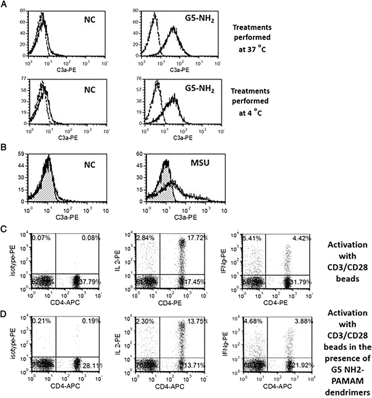
Figure 3. Membrane permeability underlies the mechanism of IC activation by cationic PAMAM dendrimers.
PBMC from at least three healthy donor volunteers were either untreated or treated G5 cationic PAMAM for 1 h. Percent of lymphocytes positive for the surface expression of C3a were analyzed by flow cytometry. (A) The experiment was performed under 37 °C or 4 °C. Dotted line – isotype; solid line – C3adesArg (B) Monosodium urate crystals (MSU) at concentration 0.1 mg/mL were tested to verify the role of other danger signals with membrane permeabilizing activity on the IC activation. Hatched histogram – isotype control; clear histogram – C3adesArg (C) T-cell activation with CD3/CD28 beads results in expression of intracellular cytokines IL-2 and IFNγ (D) The presence of C3a inducing G5 amine-terminated PAMAM dendrimers during activation with CD3/CD28 beads did not interfere with T-cell functionality. Shown are representative images from one donor. Total of at least three donors was used in each experiment, and all treatments in individual donors were tested in duplicate.

