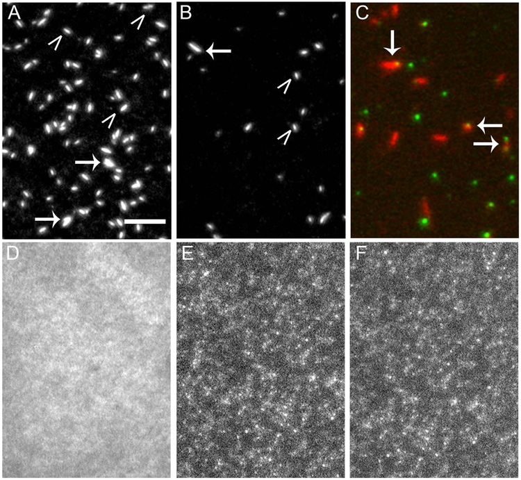
Figure 2. QD labeled RLC interferes with SMM filament formation.
TIRF microscopy images of rhodamine labeled SMM filaments in filament buffer on a glass coverslip. Images show how various modifications to the RLC interfere with filament formation. Scale bar = 5 µm and applies to all images. A) Control SMM. B) SMM with a 525 nm QD- RLC. In A and B, SMM is 20 µg ml−1, white carets point to single filaments, arrows point to aggregates. C) Overlaid image of 20 µg ml−1 SMM filaments (red) with 525 nm QD (green) chemically coupled to the RLC at Cys-108. Arrows point to co-localized filaments and QDs. D) SMM with a 585 nm QD-RLC. E) SMM with a 655 nm QD-RLC. In C and D, SMM is 0.5 µg ml−1.

