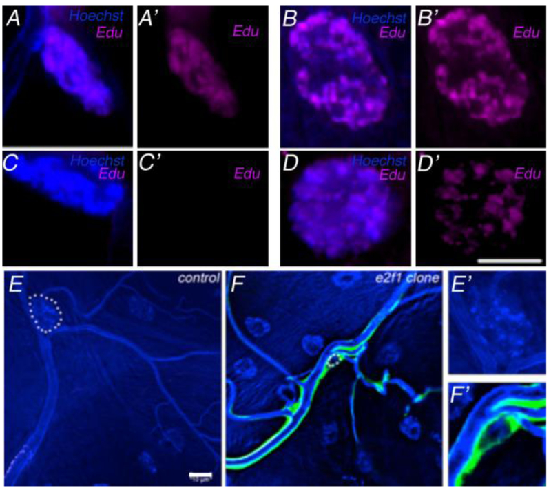
Figure 1: Endoreplication of Tracheal Terminal Cells.
Terminal cells (A, C) and muscle cells (B, D) whose nuclei were visualized by Hoechst staining (blue), were examined for incorporation of Edu (magenta) subsequent to feeding during 2nd (A,B) or early 3rd (C,D) larval instars. All larvae were scored at third wandering instar regardless of the time of feeding. Merged images are shown in A-D, Edu staining only is displayed in A’-D’). Nuclear size is dependent on endoreplication, as cells mutant for e2f1 show dramatically smaller nuclei (compare E, F). In mosaic larvae stained with Hoechst, control terminal cell nuclei (E) were much larger than e2f1 clones marked by GFP expression (F). Terminal cell nuclei are outlined in white dashed circles. Autofluorescence in the UV channel also mark the tubes of the tracheal system. (E’) and (F’) show enlarged nuclei from (E) and (F), respectively. Scale bars =10 μm.

