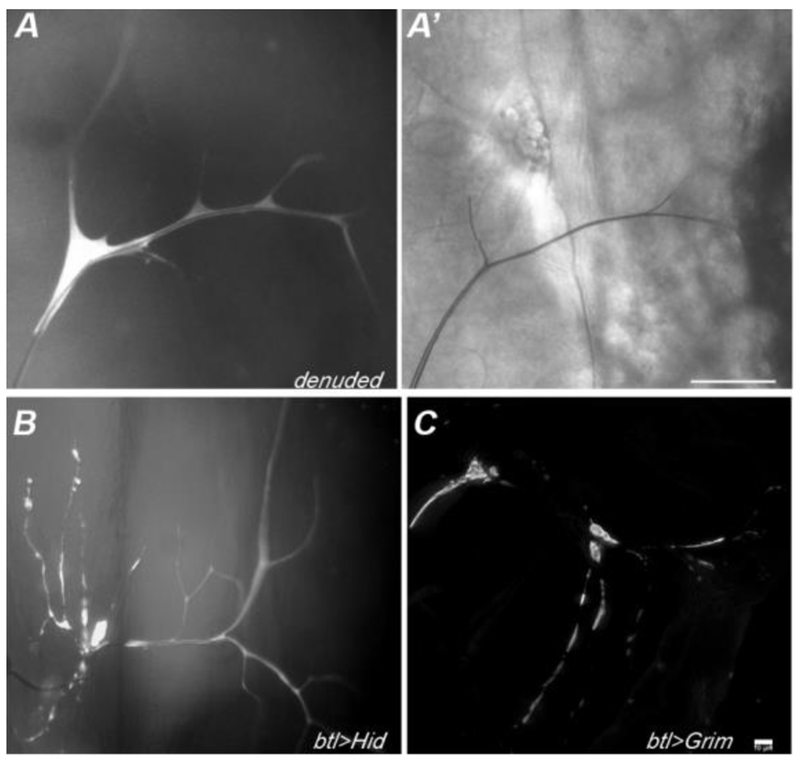
Figure 6: Terminal cells mutant for denuded show reduced size and branch complexity.
In mosaic larvae, terminal cells homozygous for denuded (15-20 allele shown) have reduced cell size and branch number. Terminal cell clones were identified as GFP (white) positive cells. In particular, while one or two main branches appear of normal size and gas-filled (A’), side branches are reduced in number and appear thin and whispy. The defects observed in denuded clones are not due to apoptosis, as terminal cells induced to apoptose by expression of Hid (B) or Grim (C), using the MARCM system, show blebbing of the cytoplasm and branch loss. Note that extensive branching occurs in these terminal cells prior to the onset of branch loss, this may reflect the delay in GAL4-induced expression associated with turn-over of GAL80 subsequent to clone induction. Scale bar in A’ = 50 microns, Scale bar for B,C (in C) = 10 microns.

