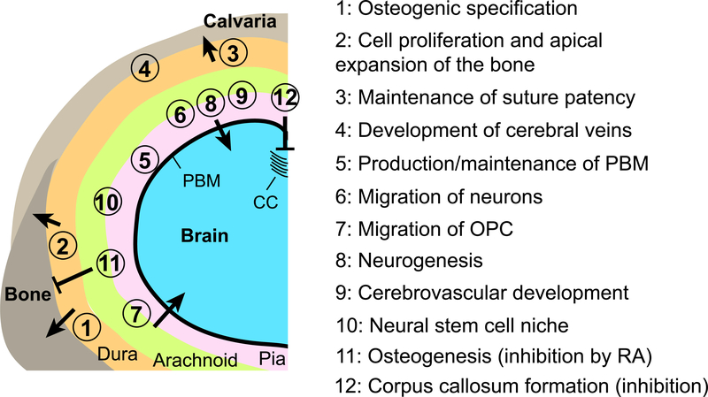
Figure 3. Influences of the meninges to development of the calvaria and the brain.
A schematic representation of a coronal section of the head of mouse embryos at late gestation stages (>E12.5). The left-dorsal quadrant of the head is shown. The positions of the circled numbers indicate the meningeal component thought to be responsible for each interaction. For the numbers straddling the arachnoid mater and the pia mater, there is not enough evidence to assign the function to only one layer or the other, and both layers are likely involved. CC: corpus callosum, OPC: oligodendrocyte precursor cells, PBM: pial basement membrane, RA: retinoic acid.

