Figure 4.
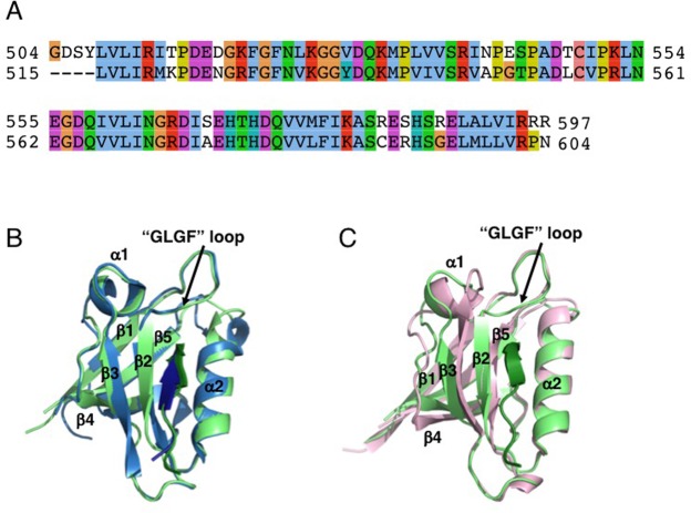
Structural comparison of the X-ray structure of PTPN3-PDZNext bound to HPV16E6 PBM with PTPN4-PDZ complexed to Cyto8retev, and with the CS-ROSETTA model of PTPN3-PDZ. (A) Clustalw2 alignment of PTPN3-PDZ (top) and PTPN4-PDZ (bottom). (B) Superposition of the structures of PTPN3-PDZNext bound to HPV16E6 PBM (light green/green) with PTPN4-PDZ bound to Cyto8retev (PDB 5EYZ) (light blue/blue). (C) Superposition of PTPN3-PDZNext bound to HPV16E6 PBM (light green/green) with the CS-ROSETTA model of PTPN3-PDZ (pink).

