Figure 4.
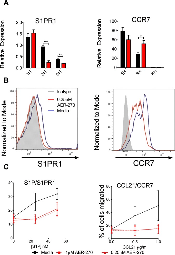
AER-270 treatment alters T cell migration toward S1P and CCL21. T cells were isolated from naive B6 mice and cultured in media only (black) or in media containing 0.25 μM AER-270 (red). (A) mRNA expression of S1PR1 and CCR7 by qRT-PCR. Gene expression was normalized against mRNA obtained from freshly isolated, untreated spleen T cells (n = 3–4, and repeated 2–3 times with similar results observed). (B) T cells were harvested after 12 h of culture, stained with antibodies against either S1PR1 or CCR7 and analyzed by flow cytometry. Representative histograms are shown (isotype control- grey, media only - blue, AER-270 - red), the experiment was repeated 2–3 times with similar results. (C) Freshly isolated T cells were stained with CFSE and 105 cells were then placed in the upper chamber of a transwell plate in media alone (black) or with AER-270 at 0.25 μM (solid red) or AER-270 at 1.0 μM (dashed red). The lower chamber contained either S1P (0–50 nM) or CCL21 (0–1 μg/ml) and the relevant concentrations of AER-270 and placed in an incubator for 90 mins at 37 °C. Cells in the lower chamber were quantified by fluorescence intensity and the results shown as percentage of cells migrated (n = 3–4, and repeated 2–3 times with similar results observed). (*p < 0.05, **p < 0.01, ***p < 0.001, no notation indicates no significant change via a one-tailed Student t-test).

