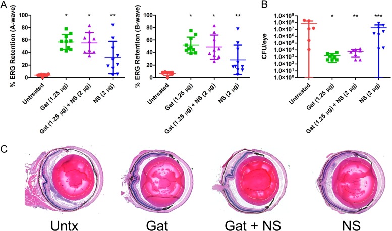
FIG 4.
Human nanosponges and human nanosponges plus gatifloxacin increased retinal function retention and protected retinal architecture following E. faecalis infection in a murine model of endophthalmitis. Right eyes of mice were infected with 100 CFU of E. faecalis. At 6 h postinfection, E. faecalis-infected right eyes were intravitreally injected with 0.5 μl PBS only (untreated control), 0.5 μl PBS containing 1.25 μg gatifloxacin (Gat), 0.5 μl PBS containing 1.25 μg gatifloxacin and 2 μg of human nanosponges (Gat+NS), or 0.5 μl PBS containing 2 μg of human nanosponges (NS). (A) Retinal function was assessed by electroretinography 24 h postinfection. Values represent means ± standard deviations (SDs) from at least 6 eyes per group in two independent experiments (A-wave: *, P < 0.0001; **, P = 0.0014; B-wave: *, P < 0.0001; **, P = 0.0021 versus untreated controls). (B) Eyes were harvested from mice and E. faecalis CFU/eye was determined. Values represent means ± SDs from at least 6 eyes per group in two independent experiments (*, P = 0.0007; **, P = 0.0013; ***, P = 0.5728 versus untreated controls). (C) Histological analysis of eyes infected with E. faecalis revealed retinal and corneal edema and cellular infiltration and fibrin deposition in both the anterior and posterior chambers; partial disruption of retinal layers was also apparent. In eyes treated with Gat and Gat+NS, retinal dissolution and edema, cellular infiltration, and fibrin deposition were reduced relative to that in untreated eyes. Eyes treated with NS only showed intact retinal layers, but cellular infiltrates and fibrin were not reduced in the anterior and posterior segments relative to that in untreated eyes.

