FIG 5.
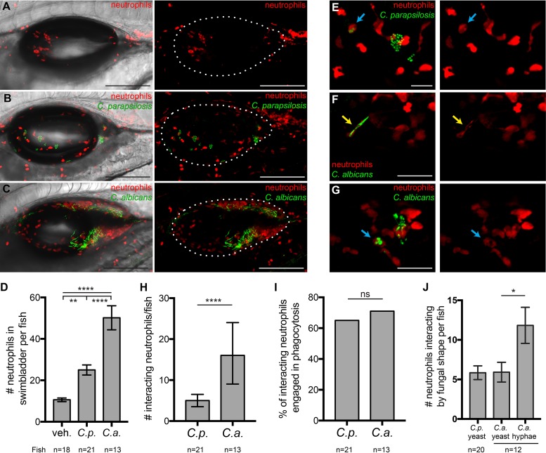
Neutrophils respond to infections with both Candida species. Tg(mpx:mCherry):uwm7Tg zebrafish (red neutrophils) were infected as described in the legend of Fig. 1 and imaged at 24 hpi. Data are pooled from 5 independent experiments. (A to C) Representative images from vehicle (A), C. parapsilosis (B), and C. albicans (C) cohorts. Maximum projections of 19 z-slices (A), 18 z-slices (B), and 16 z-slices (C), with (left) and without (right) a single DIC z-slice, are shown. (D) Neutrophils per fish in the swimbladder lumen at 24 hpi. (E to G) Examples of neutrophils (red) interacting with C. parapsilosis (green) (E) or C. albicans (green) (F and G). Interactions include contact, phagocytosis (E and G, blue arrows), and “frustrated phagocytosis” (F, yellow arrows). Maximum projections of 3 slices (E and F) and 9 slices (G) are shown. (H) Numbers of neutrophils per fish involved in interactions with C. parapsilosis or C. albicans at 24 hpi. (I) Percentages of interacting neutrophils engaged in phagocytosis at 24 hpi. (J) Numbers of neutrophils per fish interacting with yeast of C. parapsilosis and yeast or hyphae of C. albicans. Numbers of neutrophils scored for the vehicle, C. parapsilosis, and C. albicans were 191, 525, and 652, respectively. Statistics are described in Materials and Methods (*, P ≤ 0.05; **, P ≤ 0.01; ***, P ≤ 0.001; ****, P ≤ 0.0001; ns, not significant [P > 0.05]). Bars, 150 μm (A to C) and 40 μm (E to G).

