Figure 13.
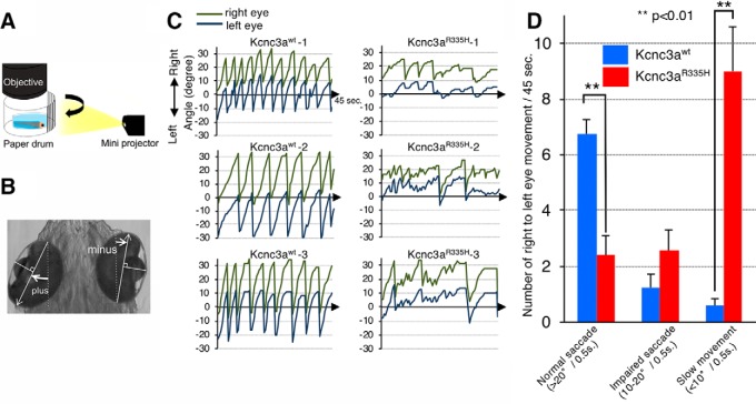
Expression of Kcnc3aR335H pathological variant in PCs causes eye movement deficits. A, Schematic drawing of the experimental set up for OKR monitoring. Moving black and white stripes from left to right were projected onto a white paper drum while the larva was immobilized in agarose with only its eyes allowed to move freely. B, Measurement of eye position. The angle of each eye was measured with respect to the external vertical axis parallel to the midline; angles of eye position deflected counterclockwise were defined as positive values, whereas clockwise deflected ones were defined as negative values. White arrows, The long axis of an ellipse; dotted line, external vertical axis. C, The graphs show left and right eye position tracking by angular eye displacement during visually triggered OKR from three representative 6.3 dpf larvae expressing Kcnc3awt (left) or Kcnc3aR335H (right) in PCs. D, Each bar in the graph indicates the average number of saccades observed during individual OKR trials (45 s) for Kcnc3awt (blue) or Kcnc3aR335H (red) larvae. Normal saccades (angle change larger than 20°/0.5 s right to left); impaired saccades (angle change at 10–20°/0.5 s right to left); or slow eye movements (angle change <10°/0.5 s right to left) were distinguished. N values for D are 6 OKR responses corresponding to 12 eye movements for 6 zebrafish larvae each. p values were determined by one-way ANOVA with post hoc Tukey HSD test. D, Kcnc3awt versus Kcnc3aR335H (Normal saccade), p = 0.0010053; Kcnc3awt versus Kcnc3aR335H (Slow movement), p = 0.0010053.

