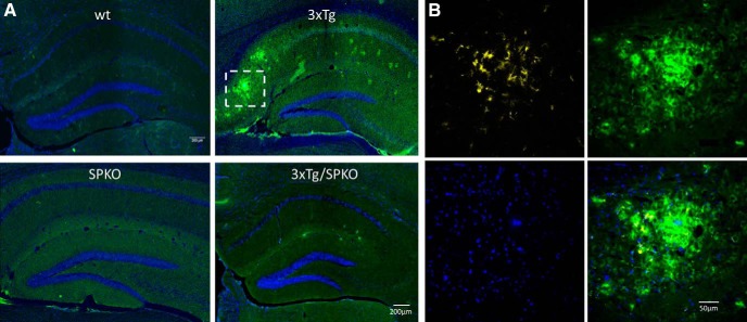
Figure 11.
β-Amyloid plaques detected in the 3xTg: Representative immunostaining of hippocampal sections. A, Immunolabeled for β-amyloid (green) and DAPI (blue). B, Enlargement of representative β-amyloid plaque colocalized with activated microglia (yellow). Nine-month-old female mice; n = 18 sections taken from 3 animals/group.

