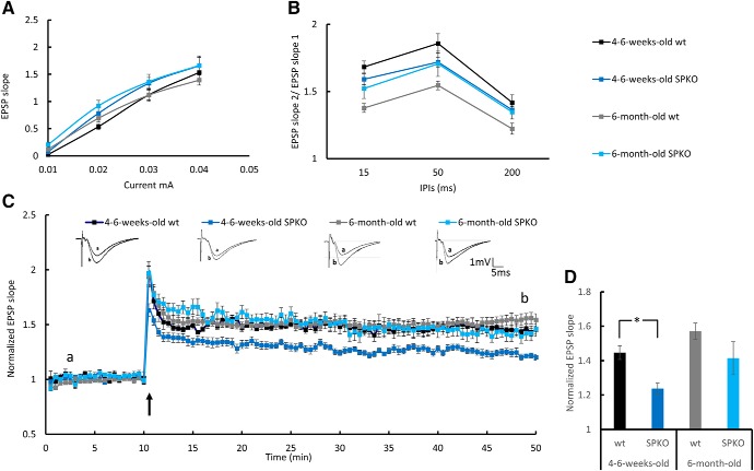
Figure 4.
Young but not adult SPKO mice show LTP deficiency: extracellular field potential recordings of EPSP in CA1 stratum radiatum of the mouse hippocampus. A, Input–output curves showing EPSP slopes recorded at different stimulation intensities. B, Paired-pulse ratio at three different interpulse intervals (IPIs). C, Normalized EPSP slopes recorded in response to tetanic stimulation. HFS was delivered to S1, marked by a black arrow. Sample EPSPs recorded before and after HFS at the time indicated in the record are presented above. D, Bar graph summarizes LTP results at time point “b” showing a significant reduction in LTP in the young SPKO group compared with the wt group (wt: n = 7 slices, 1.45 ± 0.04; SPKO: n = 7 slices, 1.24 ± 0.0; p < 0.003, two-sample t test) that was restored in the adult SPKO group. One-month-old male mice (n = 6 slices taken from 3 mice per group) and 6-month-old male mice (n = 9–15 slices taken from 3–4 mice/group). The 6-month-old group is the same cohort of results as presented in Figure 8. *p < 0.05.

