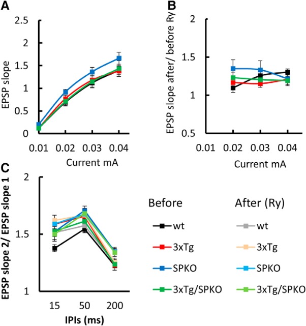
Figure 8.
3xTg/SPKO mouse line exhibits no differences in tissue viability and paired-pulse facilitation: extracellular field potential recordings of EPSPs in Schaffer collateral of the hippocampus. A, Input–output curves showing EPSP slopes recorded at different stimulation intensities. B, EPSP slope after/before 10 min of 1 μm Ry perfusion. C, Paired-pulse ratio at three different interpulse intervals (IPIs) before and after 10 min of 1 μm Ry perfusion. Six-month-old male mice; n = 9–15 slices taken from 3–4 mice/group.

