Fig. 7.

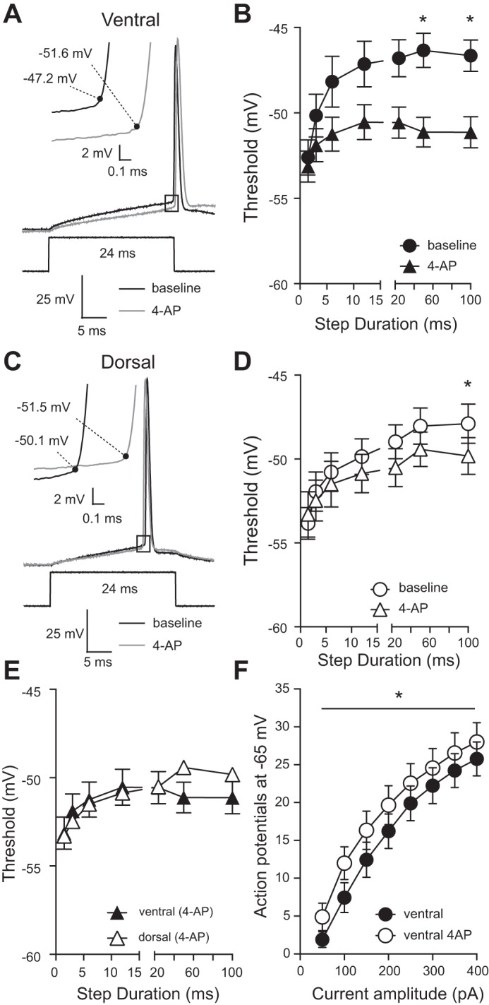
Application of 4-aminopyridine (4-AP) abolishes the difference in action potential threshold between ventral and dorsal CA1 neurons. A: representative voltage responses showing action potentials elicited from a ventral CA1 neuron before and after application of 50 µM 4-AP. Inset: expanded area indicated by the black box showing action potential threshold (black dot). B: application of 4-AP significantly hyperpolarized action potential threshold in ventral CA1 neurons (*P < 0.05) (n = 10 ventral cells from 9 mice). C: representative voltage responses showing action potentials elicited from a dorsal CA1 neuron before and after application of 50 µM 4-AP. Inset: expanded area indicated by the black box showing action potential threshold (black dot). D: application of 4-AP had no significant effect on action potential threshold in dorsal CA1 neurons (n = 7 dorsal cells from 9 mice). E: in the presence of 4-AP action potential threshold is not significantly different between dorsal and ventral CA1 neurons (n = 10 ventral cells and 7 dorsal cells from 9 mice). F: the mean number of action potentials fired during 1-s current injections of varying amplitudes differed at all current amplitudes in the presence of 4-AP in ventral CA1 neurons. Ventral: n = 9; dorsal: n = 8; n = 9 animals. Data presented as means ± SE and analyzed using 2-way ANOVA with Sidak’s multiple-comparison test. See Supplemental Table S1 for details.
