
Fig. 3.
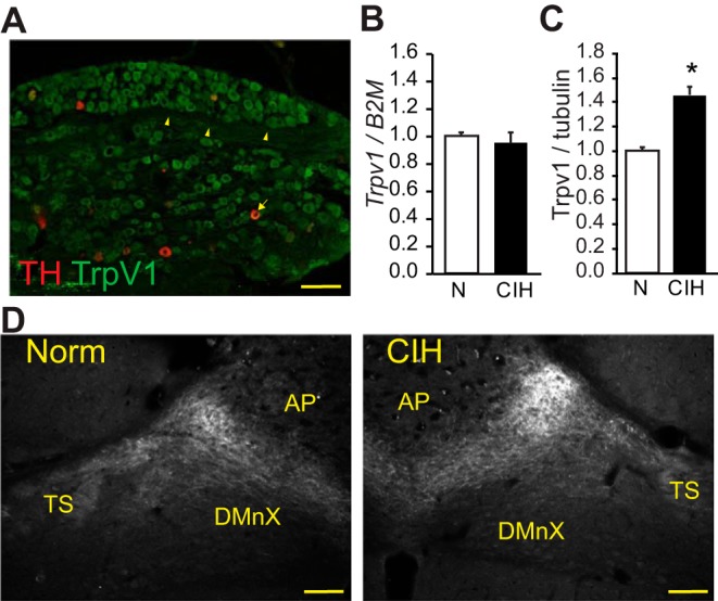
TRPV1 in sensory afferents and nucleus of the solitary tract (nTS) after chronic intermittent hypoxia (CIH). A: TRPV1 is localized in normoxic (NORM) sensory afferents of the nodose-petrosal ganglia (NPG; green) and colocalizes with tyrosine hydroxylase (TH; red). Arrowheads indicate TRPV1 fibers in NPG; arrows indicate the TRPV1 and TH co-labeled neuron. B: relative expression of TRPV1 mRNA in NPG from NORM and CIH-exposed rats (n = 3 each), as determined by the 2ΔΔCT method. Trpv1 was normalized to the housekeeping gene B2M. CIH did not alter Trpv1 mRNA. C: immunoblot analysis of TRPV1 protein from NPG tissue from NORM and CIH-exposed rats (n = 2–3). TRPV1 was normalized to tubulin. CIH elevated TRPV1 protein in NPG. *P < 0.05, t-test. D: expression of TRPV1 in the nTS of NORM and CIH-exposed rats. Note the expression of TRPV1 in the afferent-containing tractus solitarii (TS) and the tendency for increased immunofluorescence in CIH rats. Measurements were taken from 100 × 100-mm box adjacent to the TS. Bregma level ~14.04 mm. AP, area postrema; DMnX, dorsal motor nucleus of the vagus. Scale bars, 100 μm.