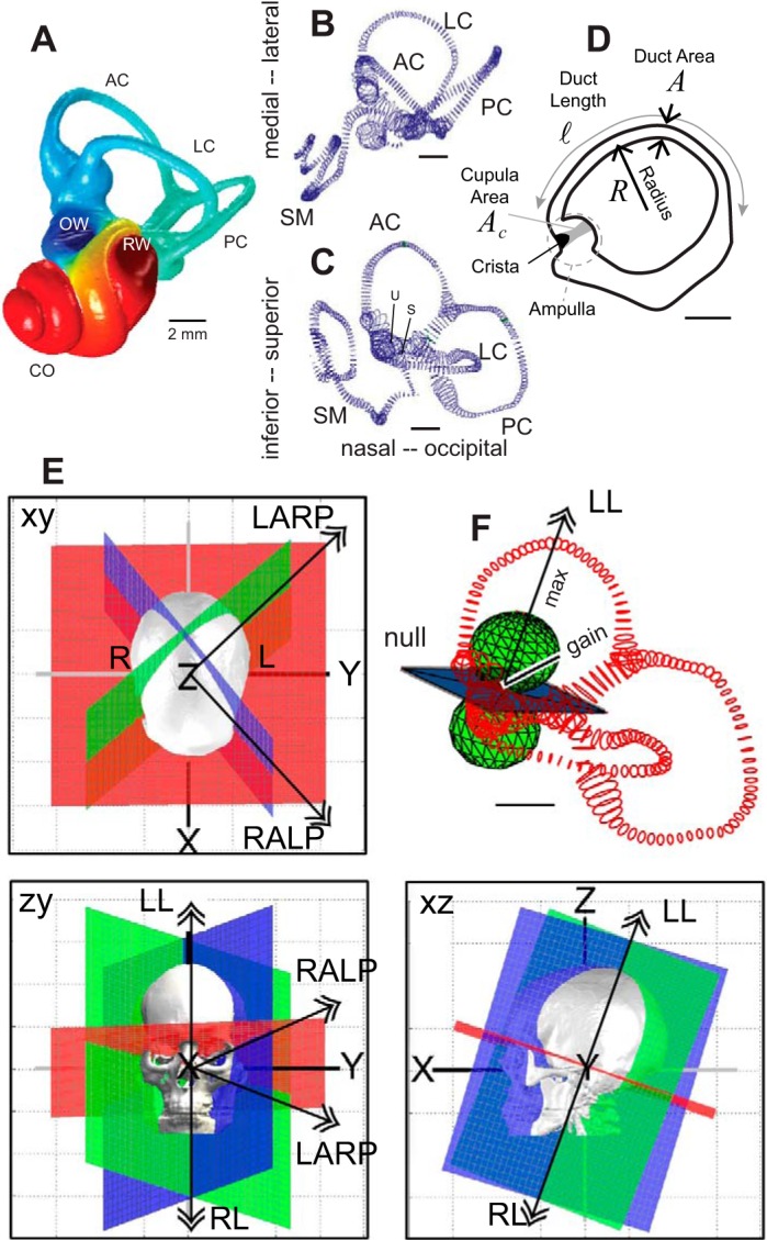
Fig. 1.

Human vestibular labyrinth. A: reconstruction of a human osseous labyrinth showing the gross morphology of the bony cavity occupied by the anterior semicircular canal (AC; blue), lateral canal (LC), posterior canal (PC; teal), and the cochlea (CO; red). The locations of the oval window (OW) and round window (RW) are indicated. B and C: adult human membranous labyrinth showing the endolymph-filled LC, AC, PC, cochlear scala media (SM), and locations of the utriculus (U) and sacculus (S) from 2 orthographic views. D: outline of the LC membranous labyrinth projected in the plane of the canal showing the location of the ampulla, cupula, crista, and cupula. Key dimensions are indicated in italics. E: anatomical canal planes determined from human computed tomography scans annotated with double arrows showing right-hand-rule rotation directions that maximize excitation of the left AC and right PC (LARP), right AC and left PC (RALP), right LC (RL), and left LC (LL). F: the mechanical gain is zero for rotation in the null plane and maximum for rotation in directions perpendicular to the null plane, shown for the LC (LL, double arrow). Sensitivity obeys the 3-dimensional cosine rule, with the magnitude of the mechanical response denoted by the distance from the pole of the sphere to the surface of the sphere (gain). [A: provided by Iversen et al. (2016); B–D: adapted from Ifediba et al. (2007); and E: adapted from Della Santina et al. (2005).]
