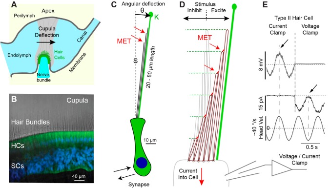
Fig. 2.
Mechanotransduction. A: schematic showing the cupula (gray) spanning the entire lumen of the ampulla and enveloping hair bundles at the surface of the sensory epithelium. B: hair cell (HC) stereocilia project as stiff actin core rods 20–80 µm into the cupula (toadfish crista. Green: xyloside (BX) in HCs and supporting cells (SCs). Blue: DAPI nuclear stain. C: schematic of type I murine HC and bundle illustrating the glycocalyx (green) around the kinocilia (K) that integrates with the cupula, and putative locations of mechanoelectrical transduction channels (MET; red arrows) at the tips of stereocilia (S) 20–80 µm above the HC body (sketch to scale). D: stereocilia are stiff and deflect almost as rigid rods pivoting at their base. Deflection toward the kinocilia increases the transduction current and is excitatory, while deflection away decreases the transduction current and is inhibitory (aspect ratio not to scale, bundles are longer than depicted). E: current- and voltage-clamp recordings of semicircular canal HC in vivo demonstrate the MET current closely follows bundle displacement for small physiological stimuli. Nonlinearities arise at higher stimulus levels. [B: provided by Holman et al. (2016); and E: adapted from Rabbitt et al. (2005).]

